CBCT Cone Beam
High-precision three-dimensional dental diagnostics. We visualize what conventional X-rays cannot show, with minimal radiation exposure.
What is CBCT?
CBCT (Cone Beam Computed Tomography) is a three-dimensional imaging technique specifically designed for the maxillofacial and dental area.
Unlike conventional X-rays that offer a flat (2D) image, CBCT generates a complete volumetric model of your teeth, bones, nerves, and tissues. It’s like going from a photograph to a hologram.
True Volumetric Imaging
The CBCT captures hundreds of images in a single 360° rotation, reconstructing a navigable 3D model that allows us to view any structure from any angle.

2D X-Ray vs 3D CBCT
2D X-Ray
Conventional Panoramic
- Flat image with structure superposition
- Needs 30-50% bone loss to detect lesions
- Does not show hidden cavitations
- Imprecise measurements due to distortion
- Does not show exact relation to nerves and sinuses
3D CBCT
Cone Beam Tomography
- Navigable 3D image without superpositions
- Detects incipient lesions and density changes
- Visualizes hidden cavitations (FDOJ)
- Exact millimetric measurements (isotropic voxel)
- Precise localization of nerves and structures
What do we use CBCT for?
In biological dentistry, CBCT is essential to see beyond the obvious and diagnose with precision.
Cavitation Detection
We identify FDOJ lesions and osteonecrosis that conventional X-rays cannot show. It is the key tool for this diagnosis.
Implant Planning
We evaluate bone density, proximity to nerves and sinuses, and plan the exact position of the implant using guided surgery.
Temporomandibular Joint (TMJ)
Diagnosis of joint bone changes: arthritis, erosions, osteofitos. For complete evaluation of soft tissues and articular disc, it is complemented with MRI.
Sinus Lift
We accurately measure available bone height and plan the most appropriate lift technique. The final plan includes clinical assessment of mucosal thickness and sinus status.
Complex Extractions
We locate the exact position of wisdom teeth, residual roots, and their relationship with nerve structures.
Pathology and Cysts
We detect, measure, and evaluate cysts, tumors, granulomas, and other pathologies with three-dimensional precision.
Why choose CBCT?
Less Radiation
Up to 6 times less radiation than a conventional medical CT. Safe for frequent dental use.
Fast Scan
The complete scan takes less than 20 seconds. Comfortable, quick, and without claustrophobia.
Sub-millimeter Precision
Resolution up to 0.1mm thanks to isotropic voxels. Exact measurements in any plane.
Complete Vision
3D navigation in all planes: axial, sagittal, coronal, and transversal. No blind spots.
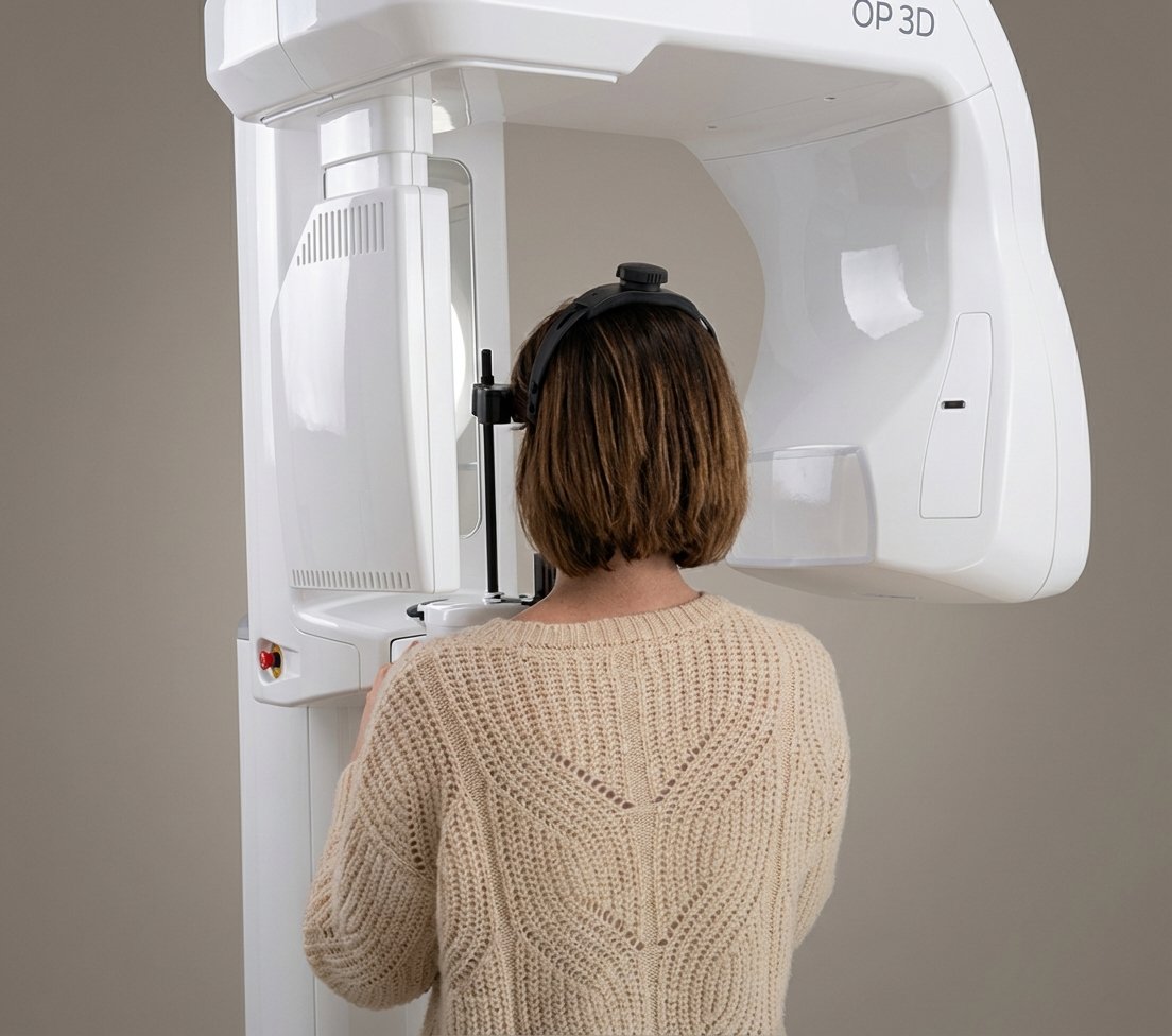
What is it like to get a CBCT?
Positioning
You stand in front of the machine. We support your chin and forehead to stabilize.
30 secondsScanning
The arm rotates 360° around your head capturing hundreds of images.
10-20 secondsReconstruction
The software reconstructs the complete 3D model of your maxillofacial structure.
1-2 minutesDiagnosis
Your dentist analyzes the images and explains the findings to you right there.
ImmediateImportant Clarification
CBCT is a high-precision diagnostic support tool that requires clinical correlation. At BDS, all reports include:
- Interpretation by a qualified professional in dental radiology
- Clinical evaluation and review of patient history
- Complementary tests when indicated
- Recommendations based on comprehensive assessment
Note: CBCT radiation dose varies according to Field of View (FOV) and acquisition parameters. Its use is avoided during pregnancy unless clinically justified. This service complements, but does not replace, in-person medical/dental assessment.
Frequently Asked Questions
The dose varies depending on the scanned area. Limited scan: similar to a panoramic X-ray. Full scan: 0.1-0.3 mSv, still significantly lower than a medical CT (2.5 mSv).
No, it is completely painless and very comfortable. You simply stand (or sit) in front of the machine for a few seconds while the arm rotates around you. There are no closed tubes or feelings of claustrophobia. You just have to stay still for 10-20 seconds.
The entire process takes approximately 5 minutes, and the diagnosis is immediate. Once the scan is done, the images are processed in 1-2 minutes and your dentist can review them with you right there, explaining the findings on the screen.
A panoramic X-ray is a flat 2D image where structures overlap. CBCT offers a true 3D image where we can navigate layer by layer, measure with millimetric precision, and detect lesions that would remain hidden in 2D. For detecting cavitations, for example, CBCT is essential.
As a general rule, X-rays are avoided during pregnancy unless strictly necessary. Although CBCT radiation is low, we prefer to postpone non-urgent diagnostic tests until after pregnancy. If you have questions, consult us and we will evaluate your individual case.
Other Treatments
Zirconia Implants
100% biocompatible
See moreAmalgam Removal
SMART Protocol
See moreFDOJ Cavitations
Hidden inflammation
See moreDental Ozone Therapy
Natural disinfection
See moreRegenerative PRF
Your own blood
See moreIntegrative Orthodontics
Dr. Patricia Díaz
See moreBiological Pediatric Dentistry
Dr. Brenda Martínez
See moreIntegrative Medicine
Dr. Patrick Welter
See morePrecision Diagnosis
Discover what conventional X-rays cannot show. A 3D diagnosis can completely change your treatment approach.